|
Ian Brown Graduate Student Paper of the Year:
The concept of phagocytosis is understood as the process by which white blood cells in your immune system “eat” potentially harmful exogenous agents. After all, the term phagocytosis is comprised of the Greek words phago and cyto, meaning “eating” and “cell” respectively. The “eating” portion specifically implies “engulfment” when referring to phagocytosis. What if it turns out that “eating” is an incomplete metaphor for this important immune response? Our research demonstrates that white blood cells in your body also have the capability to “chew” its phagocytic content, as opposed to only “swallowing” it whole. Of note, we feel it appropriate to anthropomorphize these concepts to communicate their essence. Enter a process coined F-actin flashing. It is a peculiar case of seemingly spontaneous and repeated polymerization of filamentous actin (or F-actin) around phagosomes in white blood cells. Such a cellular event has been observed and described in the recent past, but its mechanism and role had been left unexplained. Our research helps address these gaps in our understanding. We were able to determine that this process has a particular cadence of repeated polymerization of F-actin on phagosomes and has a higher incidence in complement receptor 3 (CR3)-mediated phagocytosis, one of the two main phagocytic pathways. Furthermore, we determined that accompanying F-actin are myosin and mechanosensitive proteins typically present in focal adhesions. Actomyosin provides the contractile force applied on phagosomes, and the cascade of mechanosensitive proteins modulates the cadence of F-actin flashing depending on the stiffness of the engulfed particle. Notably, we found that the pressure applied on phagosomes by the flurries of actomyosin contractions – the “chewing” – break down engulfed red blood cells into smaller pieces, effectively increasing the surface area available for proteolytic enzymes to act upon. This process accelerates phagocytic content degradation, and in turn, phagosome maturation. This finding represents a newly discovered tool for macrophages. To summarize, F-actin flashing is a process by which actomyosin and mechanosensitive proteins repeatedly assemble and contract around phagosomes, akin to chewing, breaking its content into smaller constituents. Ultimately, those remaining pieces will be degraded at a faster rate, ridding the macrophage of the opsonized particle. It may be revealed by further research that F-actin flashing may be directly involved with antigen presentation, a critical step in the process of developing acquired immunity.
|
|
Ecology & Evolution Graduate Student Paper of the Year:
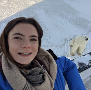
High levels of disease often persist in wildlife populations, but the complexity and diversity of parasite life cycles makes determining infection pathways challenging. This complexity compromises our ability to predict how infection dynamics might be influenced by anthropogenic disturbances, which, in turn, affects our ability to mitigate negative impacts on human and wildlife hosts. Mathematical models of host‐parasite interactions are powerful tools for exploring complex ecological interactions and allow researchers to formally test the plausibility of hypothesized mechanisms, even when empirical data seem scarce. Despite the broad applicability of this approach, the full potential of mathematical models to address fundamental problems in ecology with minimal data remains underappreciated. Our manuscript demonstrates the utility of a modelling framework for addressing a decades‐long controversy regarding the source of Trichinella nativa infections in polar bears. We find strong support that consumption of infected seals can produce the high levels of T. nativa that are observed in the Southern Beaufort Sea (SBS) polar bear population and elsewhere, and we find no support for the historically preferred hypotheses invoking scavenging, cannibalism, and conspecific scavenging as key transmission pathways. Seals had previously been largely dismissed as a source of T. nativa infection for polar bears due to the extremely low prevalence observed in seal populations, but our models demonstrate that the polar bear’s specialized seal diet nevertheless results in a high cumulative infection risk over a bear’s lifetime. Our results highlight how minimal data, such as the prevalence of parasitic infections in a population, combined with mechanistic models of the transmission dynamics, can clarify complex, unobservable interactions and challenge prevailing assumptions. |
|
Postdoc Paper of the Year:
Infectious disease is an increasing threat to wildlife due to, for example, the emergence and spread of new parasites associated with climate change and more frequent contact with domesticated animals. Migratory animals are particularly vulnerable because their long-distance journeys expose them to a variety of habitats and hosts, facilitating transmission and enabling parasite range expansions. Understanding infectious disease in migratory animals is challenging because the vast distances covered result in variable host densities and infection pressures over space and time, thus also making it difficult to collect data that allow disentangling the underlying transmission mechanisms and dynamics. Empirical studies show that migrants may have higher, lower, or the same infection intensity as resident populations, and a variety of mechanisms have been proposed to explain such seemingly idiosyncratic observations. In order to unravel the mechanisms leading to these outcomes and assess likely impacts of global change on wildlife health and infectious disease risk, process-based models are needed that explicitly account for the spatiotemporal dynamics of host-parasite interactions. In this article, we present such a model and use it to understand the general conditions that improve host population health via migratory escape and recovery from parasitism, or via the migratory culling or stalling of infected individuals. Simulations show that different infection patterns and migration outcomes arise from different model parameterizations, depending on characteristics of the host, the parasite, and the environment – thus unifying migratory escape, migratory culling, and migratory stalling within a common mechanistic framework, and laying a theoretical foundation for exploring what may be driving the diverse patterns observed in nature. While previous theory has focused on specific cases, usually involving microparasites where the intensity of infection is ignored, our framework also allows tracking the variance in parasite burdens among hosts. This provides an explicit link to empirical studies, suggesting when and where data need to be collected in order to distinguish mechanisms and inform targeted management and conservation efforts. Finally, there is increasing recognition of the interconnectedness of environmental, human, and animal health, and the roles that wildlife can play in affecting the health of people (and vice versa). Migratory wildlife in particular can connect disparate habitats and populations, potentially facilitating the spread of emerging zoonotic diseases. Frameworks, such as the one we present, for understanding and predicting disease risk among wildlife hosts – and potential risks to humans – are needed now more than ever. |


